
Search
- Page Path
- HOME > Search
Erratum
- Miscellaneous
- Erratum: Correction of Figures. Association between the Blautia/Bacteroides Ratio and Altered Body Mass Index after Bariatric Surgery
- Yoonhong Kim, Dooheon Son, Bu Kyung Kim, Ki Hyun Kim, Kyung Won Seo, Kyoungwon Jung, Seun Ja Park, Sanghyun Lim, Jae Hyun Kim
- Endocrinol Metab. 2022;37(4):701-702. Published online July 21, 2022
- DOI: https://doi.org/10.3803/EnM.2022.401
- Corrects: Endocrinol Metab 2022;37(3):475

- 1,766 View
- 80 Download

Original Articles
- Diabetes, Obesity and Metabolism
- Association between the Blautia/Bacteroides Ratio and Altered Body Mass Index after Bariatric Surgery
- Yoonhong Kim, Dooheon Son, Bu Kyung Kim, Ki Hyun Kim, Kyung Won Seo, Kyoungwon Jung, Seun Ja Park, Sanghyun Lim, Jae Hyun Kim
- Endocrinol Metab. 2022;37(3):475-486. Published online June 29, 2022
- DOI: https://doi.org/10.3803/EnM.2022.1481
- Correction in: Endocrinol Metab 2022;37(4):701
- 3,160 View
- 124 Download
- 7 Web of Science
- 7 Crossref
-
 Abstract
 PDF
PDF Supplementary Material
 PubReader
  ePub
ePub - Background
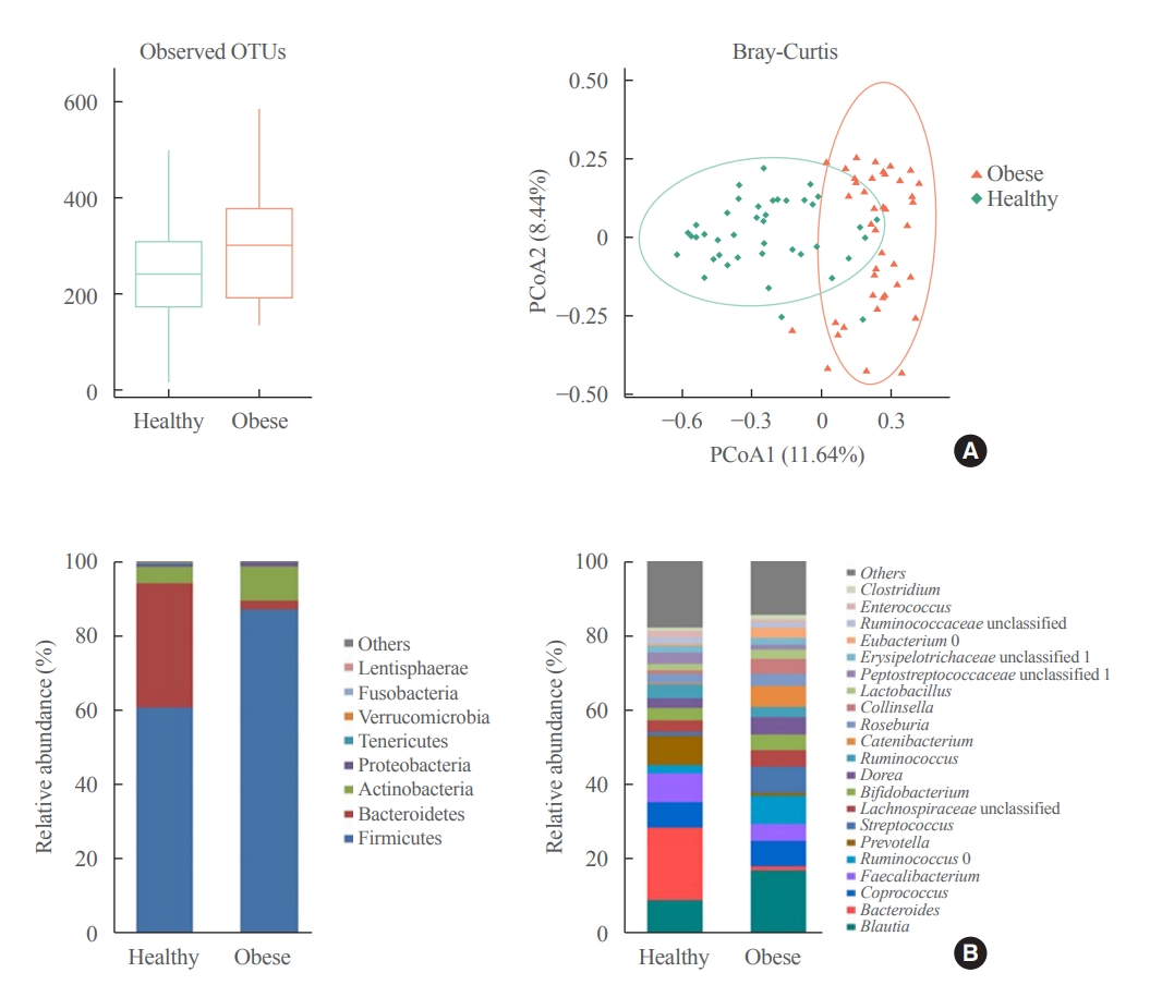
Current evidence support that the gut microbiota plays a potential role in obesity. Bariatric surgery can reduce excess weight and decrease the risk of life-threatening weight-related health problems and may also influence gut microbiota. In this study, we aimed to investigate the changes in gut microbiota before and after bariatric surgery and evaluate the association of the gut microbial shift and altered body mass index (BMI) after bariatric surgery.
Methods
Between January 2019 and July 2020, stools from 58 patients scheduled for bariatric surgery were collected. Six months after bariatric surgery, stools from 22 of these patients were re-collected, and the changes in gut microbiota before and after bariatric surgery were evaluated. In addition, the differences in gut microbiota between patients with severe obesity (BMI >35 kg/m2, n=42) and healthy volunteers with normal BMI (18.8 to 22.8 kg/m2, n=41) were investigated.
Results
The gut microbiota of patients who underwent bariatric surgery showed increased α-diversity and differed β-diversity compared with those before surgery. Interestingly, Blautia was decreased and Bacteriodes was increased at the genus level after bariatric surgery. Further, the Blautia/Bacteroides ratio showed a positive correlation with BMI. To validate these results, we compared the gut microbiota from severely obese patients with high BMI with those from healthy volunteers and demonstrated that the Blautia/Bacteroides ratio correlated positively with BMI.
Conclusion
In the gut microbial analysis of patients who underwent bariatric surgery, we presented that the Blautia/Bacteroides ratio had changed after bariatric surgery and showed a positive correlation with BMI. -
Citations
Citations to this article as recorded by
- Modulation of the gut microbiome and Firmicutes phylum reduction by a nutraceutical blend in the obesity mouse model and overweight humans: A double‐blind clinical trial
Victor Nehmi‐Filho, Jessica Alves de Freitas, Lucas Augusto Franco, Roberta Cristina Martins, José Antônio Orellana Turri, Aline Boveto Santamarina, Joyce Vanessa da Silva Fonseca, Ester Cerdeira Sabino, Bruna Carvalho Moraes, Erica Souza, Gilson Masahiro
Food Science & Nutrition.2024; 12(4): 2436. CrossRef - Natural emulsifiers lecithins preserve gut microbiota diversity in relation with specific faecal lipids in high fat-fed mice
Chloé Robert, Armelle Penhoat, Leslie Couëdelo, Magali Monnoye, Dominique Rainteau, Emmanuelle Meugnier, Sofia Bary, Hélène Abrous, Emmanuelle Loizon, Pranvera Krasniqi, Stéphanie Chanon, Aurélie Vieille-Marchiset, François Caillet, Sabine Danthine, Huber
Journal of Functional Foods.2023; 105: 105540. CrossRef - Effects and action mechanisms of lotus leaf (Nelumbo nucifera) ethanol extract on gut microbes and obesity in high-fat diet-fed rats
Zhang Yanan, Ma Lu, Zhang Lu, Huo Jinhai, Wang Weiming
Frontiers in Nutrition.2023;[Epub] CrossRef - First characterization of the intestinal microbiota in healthy Tunisian adults using 16S rRNA gene sequencing
Ahlem Mahjoub Khachroub, Magali Monnoye, Nour Elhouda Bouhlel, Sana Azaiez, Maha Ben Fredj, Wejdene Mansour, Philippe Gérard
FEMS Microbiology Letters.2023;[Epub] CrossRef - Gut microbiota and nonalcoholic fatty liver disease
Boyeon Kim, Bukyung Kim
Kosin Medical Journal.2023; 38(3): 169. CrossRef - Obésité et risque cardiovasculaire : le rôle de la chirurgie bariatrique dans la modulation du microbiote intestinal
Davide Masi, Mickael Massicard, Karine Clément
Nutrition Clinique et Métabolisme.2023; 37(2): 2S8. CrossRef - The Related Metabolic Diseases and Treatments of Obesity
Ming Yang, Shuai Liu, Chunye Zhang
Healthcare.2022; 10(9): 1616. CrossRef
- Modulation of the gut microbiome and Firmicutes phylum reduction by a nutraceutical blend in the obesity mouse model and overweight humans: A double‐blind clinical trial
- Hypothalamus and Pituitary Gland
- Metabolic Impacts of Discontinuation and Resumption of Recombinant Human Growth Hormone Treatment during the Transition Period in Patients with Childhood-Onset Growth Hormone Deficiency
- Yun Jeong Lee, Yunha Choi, Han-Wook Yoo, Young Ah Lee, Choong Ho Shin, Han Saem Choi, Ho-Seong Kim, Jae Hyun Kim, Jung Eun Moon, Cheol Woo Ko, Moon Bae Ahn, Byung-Kyu Suh, Jin-Ho Choi
- Endocrinol Metab. 2022;37(2):359-368. Published online April 25, 2022
- DOI: https://doi.org/10.3803/EnM.2021.1384
- 4,430 View
- 184 Download
- 3 Web of Science
- 3 Crossref
-
Abstract
PDFSupplementary MaterialPubReader ePub
- Background
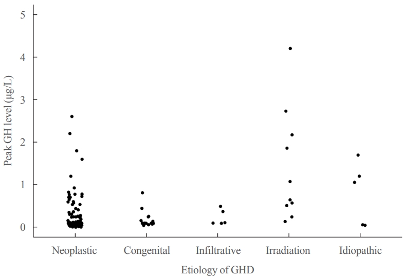
Discontinuing growth hormone (GH) treatment during the transition to adulthood has been associated with adverse health outcomes in patients with childhood-onset growth hormone deficiency (CO-GHD). This study investigated the metabolic changes associated with interrupting GH treatment in adolescents with CO-GHD during the transition period.
Methods
This study included 187 patients with CO-GHD who were confirmed to have adult GHD and were treated at six academic centers in Korea. Data on clinical parameters, including anthropometric measurements, metabolic profiles, and bone mineral density (BMD) at the end of childhood GH treatment, were collected at the time of re-evaluation for GHD and 1 year after treatment resumption.
Results
Most patients (n=182, 97.3%) had organic GHD. The median age at treatment discontinuation and re-evaluation was 15.6 and 18.7 years, respectively. The median duration of treatment interruption was 2.8 years. During treatment discontinuation, body mass index Z-scores and total cholesterol, low-density lipoprotein, and non-high-density lipoprotein (HDL) cholesterol levels increased, whereas fasting glucose levels decreased. One year after GH treatment resumption, fasting glucose levels, HDL cholesterol levels, and femoral neck BMD increased significantly. Longer GH interruption (>2 years, 60.4%) resulted in worse lipid profiles at re-evaluation. The duration of interruption was positively correlated with fasting glucose and non-HDL cholesterol levels after adjusting for covariates.
Conclusion
GH treatment interruption during the transition period resulted in worse metabolic parameters, and a longer interruption period was correlated with poorer outcomes. GH treatment should be resumed early in patients with CO-GHD during the transition period. -
Citations
Citations to this article as recorded by- Ghrelin regulating liver activity and its potential effects on liver fibrosis and Echinococcosis
Jiang Zhu, Tanfang Zhou, Meng Menggen, Kalibixiati Aimulajiang, Hao Wen
Frontiers in Cellular and Infection Microbiology.2024;[Epub] CrossRef - Relationship between the Stimulated Peak Growth Hormone Level and Metabolic Parameters in Children with Growth Hormone Deficiency
Seong Yong Lee
The Ewha Medical Journal.2023;[Epub] CrossRef - Dyslipidaemia and growth hormone deficiency – A comprehensive review
Matthias Hepprich, Fahim Ebrahimi, Emanuel Christ
Best Practice & Research Clinical Endocrinology & Metabolism.2023; 37(6): 101821. CrossRef
- Ghrelin regulating liver activity and its potential effects on liver fibrosis and Echinococcosis
- Influence of Early Age at Menopause on Bone Mineral Density and Biochemical Bone Marker.
- Young Joo Park, Chan Soo Shin, Do Joon Park, Jung Koo Kim, Sung Yeon Kim, Bo Yeon Cho, Hong Gyu Lee, Jae Hyun Kim, In Kyung Chung
- J Korean Endocr Soc. 1999;14(2):346-354. Published online January 1, 2001
- 1,159 View
- 18 Download
-
Abstract
PDF
- BACKGROUND
Among the various factors affecting bone mass and bone metabolism, aging and menopause play a major role. After the disappearance of the menstrual cycle, estrogen deficiency is the most important factor in bone loss. It is still unclear whether women with early menopause have a rate of bone loss different from women whose menopause has occurred later. Various biochemical bone markers are increased after menopause but it is still unclear whether women with early menopause have biochemical bone markers different from women whose menopause has occurred later. The aim of this study was to establish whether healthy women with early or normal menopause have different bone mass, biochemical bone markers and rates of bone loss. METHODS: Postmenopausal healthy women were divided into two groups according to their age at menopause(AAM): one group with AAM > 43 years, and the other group with AAM 50 years. Bone mass was measured using a dual energy X-ray absorptiometry(DEXA) in the lumbar, femur neck, femur trochanter, and Wards triangle. Serum levels of bone alkaline phosphatase and osteocalcin, and urine levels of calcium, deoxypyridinoline and type I collagen N-telopeptide were measured using a commercial kit. RESULTS: Age and body mass index in the early menopause group were different from those in the normal menopause group. All the bone mass and the biochemical bone markers in the early menopause group were not different from those in the normal menopause group. We selected 15 subjects from the two groups matched by age and BML Bone mass of femur neck in the early menopause group was lower than in the normal menopause group matched by age and BMI. Bone mass in lumbar, femur trochanter, and Wards triangle was lower in the early menopause group than in the normal menopause group, but the difference between the two groups was not significant. After adjusting years since menopause, we didnt find the difference of bone mass between the two groups. All the bone biochemical markers were not different in the two groups matched by age and BMI. CONCLUSION: Our data suggest that women with early menopause dont lose bone faster than women with normal menopause.
 First
First Prev
Prev


